By Ashwini Sakharkar 16 Sep, 2024
Collected at: https://www.techexplorist.com/new-technology-measure-neural-activity-babies-brains/89699/
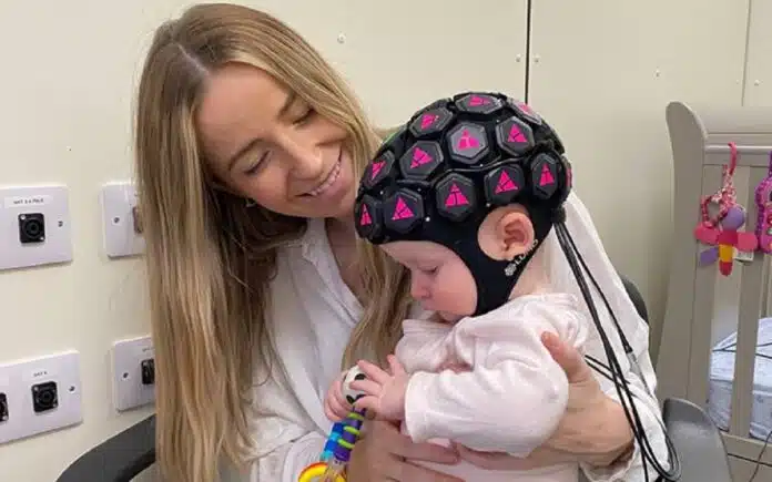
In a new study led by researchers at UCL and Birkbeck, a new technology that uses light waves to measure activity in babies’ brains has provided the most complete picture to date of functions like hearing, vision, and cognitive processing outside a conventional brain scanner.
The wearable brain imaging headgear, which was developed in collaboration with UCL spin-out Gowerlabs, found unexpected activity in the prefrontal cortex, a critical area for processing emotions, demonstrating that infants as young as five months old begin processing social stimuli.
This latest advancement allows for the measurement of neural activity across the entire outer surface of a baby’s brain, a remarkable improvement from the previous version, which could only measure activity in limited areas.
The advancement of this new technology has the potential to revolutionize our understanding of brain development, particularly in the early stages of childhood. By mapping the connections between different brain regions, we can gain valuable insights into typical and atypical neurodevelopment. This could greatly contribute to our understanding of conditions such as autism, dyslexia, and ADHD.
The findings of the early tests, along with the development of the new device, have been documented in a recent study published in Imaging Neuroscience and will be presented at the British Science Festival on Saturday 14 September.
“Previously, we developed a wearable imaging approach that could map activity in specific areas of the brain,” said Dr Liam Collins-Jones, the first author of the study at UCL Medical Physics & Biomedical Engineering and the University of Cambridge. “But this made it difficult to get a complete picture as we could only focus on one or two areas in isolation, whereas in reality, different parts of the brain work together when navigating real-world scenarios.
“The new method allows us to observe what’s happening across the whole outer brain surface underlying the scalp, which is a big step forward. It opens up possibilities to spot interactions between different areas and detects activity in areas that we might not have known to look at previously.
“This complete picture of brain activity could enhance our understanding of how the baby brain functions as it interacts with the surrounding world, which could help us optimize support for neurodiverse children early in life.”
“This is the first time that differences in activity across such a wide area of the brain have been measured in babies using a wearable device, including parts of the brain involved in processing sound, vision, and emotions,” said Professor Emily Jones, an author of the study from Birkbeck, University of London.
“The technology developed and tested in this study is a stepping stone towards a better understanding of the brain processes that underlie social development, which we haven’t been able to observe before, outside of the very restrictive bounds of an MRI scanner.
“With this, we should be able to see what’s happening in babies’ brains as they play, learn, and interact with other people in a very natural way.”
The new device underwent testing on sixteen babies aged five to seven months. While wearing the device, the babies sat on their parent’s lap and were shown videos of actors singing nursery rhymes to replicate a social scenario, and videos of moving toys, such as a ball rolling down a ramp, to replicate a non-social scenario.
Significant differences in brain activity were observed between the two scenarios. Unexpected findings in the prefrontal cortex were noted in response to social stimuli, and the researchers also found that activity was more localized in response to social stimuli compared to non-social stimuli, corroborating previous findings from optical neuroimaging and MRI studies.
Up until now, the most comprehensive method for observing brain activity has been magnetic resonance imaging (MRI), which requires the subject to lie very still within the scanner for potentially 30 minutes or more.
One limitation of traditional brain imaging methods is their inability to capture natural interactions or tasks, especially with infants who would need to be asleep or restrained for successful MRI scans.
To address this, researchers have turned to high-density diffuse optical tomography (HD-DOT) to create wearable devices that can study brain activity in a more natural setting. Not only is this technology more affordable and portable than MRI, but it also allows for a more comprehensive scan of an infant’s entire head.
The device used in the study was adapted from a commercial system developed by Gowerlabs, a UCL spin-out company established by researchers from UCL’s Biomedical Optics Research Laboratory in 2013.
“This device is a great example of academic research and commercial technological development working hand-in-hand,” said Dr Rob Cooper, senior author of the study from UCL Medical Physics & Biomedical Engineering. “The long-standing collaboration between UCL and Gowerlabs, along with our academic partners, has been fundamental to the development of wearable HD-DOT technology.”
Journal reference:
- Liam H. Collins-Jones, Louisa K. Gossé, Borja Blanco, Chiara Bulgarelli, Maheen Siddiqui, Ernesto E. Vidal-Rosas, Nida Duobaitė, Reuben W. Nixon-Hill, Greg Smith, James Skipper, Tim Sargent, Samuel Powell, Nicholas L. Everdell, Emily J.H. Jones, Robert J. Cooper. Whole-head high-density diffuse optical tomography to map infant audio-visual responses to social and non-social stimuli. Imaging Neuroscience, 2024; DOI: 10.1162/imag_a_00244
Leave a Reply